|
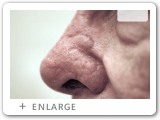
Click on an image to enlarge it and see the surgeon's comments. Follow the numbers to see the whole procedure in the right sequence. You can view the entire process in steps (click on, view and then close each image separately) or as a slide show (click on any image to view it, then use left/right arrows on your keyboard or arrows that appear on the image control bar to navigate through all images/steps). If you'd like to look at two or more enlarged images - open as many as you like and position them on your screen by moving image windows.
|
|
1.
|
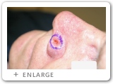
1. Boarders of the visible tumor are marked.
|
2.
|
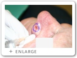
2. Surgical site is frozen with local anesthetic.
|
3.
|
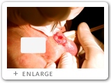
3. The resection margin is scored approximately 1-2 mm outside the visible tumor edge.
|
4.
|

4. The first resection level is removed with assistant supporting the flexible nasal rim. The surgeon bevels the resection edge to allow ease of placement during tissue processing.
|
|
5.
|
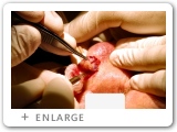
5. A deeper resection is required to remove the first level of an ulcerated or invasive cancer.
|
6.
|
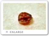
6. The tumor is oriented on the back table in the operating theatre.
|
7.
|
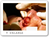
7. Bleeding is controlled with electrocautery.
|
8.
|
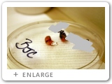
8. The cancer segment is divided into two pieces, oriented with dyes and placed in a Petrie dish labeled with the patient's name.
|
|
9.
|
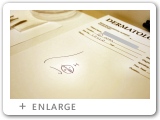
9. A schematic orientation map is made of the tissue segments to assist in reorienting the surgeon to the precise location on the patient after microscopic analysis.
|
10.
|
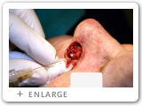
10. The surgical site is refrozen prior to removing an additional segment of tissue where cancer cells remain.
|
11.
|
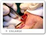
11. Removal of second tissue level with narrow margin.
|
12.
|
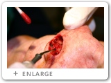
12. Bleeding is again controlled with electrocautery.
|
|
13.
|
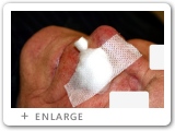
13. Example of temporary dressing used between cancer resection stages.
|
14.
|
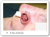
14. Surgeons examine the final defect prior to planning a reconstruction.
|
15.
|
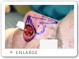
15. A local bilobed flap is designed for reconstructing of the Mohs defect.
|
16.
|
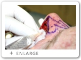
16. The surgical field is frozen with anesthetic.
|
|
17.
|
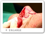
17. The edge of the flap is scored and redundant tissue excised.
|
18.
|
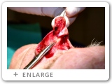
18. The nasal flaps are elevated below the nasal muscles.
|
19.
|
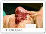
19. The flaps are inset with deep buried stitches. The skin surface is sutured with non-dissolving stitches.
|
|
|
|
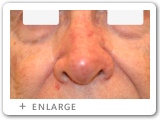
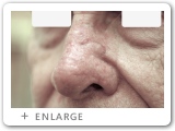
6 months following surgery:
|
|
20.
|
|
21.
|
|
22.
|
|
|
|


